• Skip to primary navigation
  • Skip to main content
  • Home
  • People
  • Research
  • Publications
  • Patents
  • Gallery
  • In the Media
  • Facilities
  • Openings

The Bioinspired Translational Microsystems Laboratory

BioinSyst

Texas A&M University College of Engineering

Research

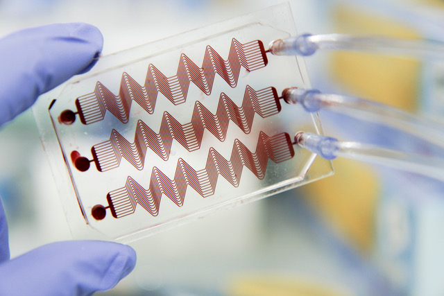
Engineering Medicine with Organ-Chips

a fast-track platform for translational research

Animals have long been the bedrock behind our understanding of human physiology and drug discovery despite the awareness of the discordance between animal and human studies. In past three decades, the number of FDA-approved drugs per billion US dollars spent on R&D has actually decreased monotonically. Our research aspires to reverse this poor trend by innovating new bioengineered approaches, paradigms and tools that make a positive impact to medicine and healthcare economics. Our lab is achieving this ambition by harnessing the basic knowledge and methods offered by cell and molecular biology, biomechanics, microfabrication technology, biomaterials, and mathematics; and reconstructing the physical microenvironment of human tissues and organs in microfluidic devices, also known as organs-on-chip. Our lab particularly specializes in making patient-specific organ-chip models of rare and orphan diseases with an emphasis on vascular and hematological diseases and its long-term manifestations in cancer, diabetes and infections. By establishing and leveraging extensive collaborations across the Texas Medical Center, our group has made contributions in advancing the fundamental understanding and drug discovery of sickle cell disease, type-1 diabetes, vein thrombosis, ovarian cancer, lymphedema and COVID-19.

Funding

Current Directions

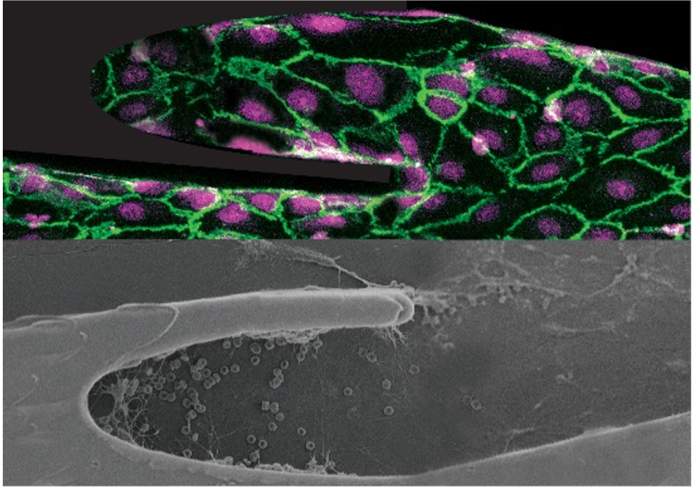
Vein-Chip: Discovery of pathophysiology & treatments of chronic venous insufficiency and thrombosis

The lack of proper models to study the formation and treatment of deep vein thrombosis (DVT) has led DVT to become one of the top three cardiovascular diseases in the world. We still don’t know what is the most effective strategy against COVID-19 induced clotting, that is shown to occur in many patients who died of it. We have leveraged our organ-on-chip methodology to create a Vein-Chip platform integrating fully vascularized venous valves and its hemodynamics, only seen in vivo prior to this work. Our Vein-Chips reveal how the vascular tissue is locally influenced by the disturbed microenvironment and depending on biological or physical cues, may either protect or exacerbate blood clot formation. The platform eventually provides a systematic approach to test and discover antithrombotic drugs and also assess COVID-19 infection or vaccination-induced clotting, which other preclinical models won’t do it.

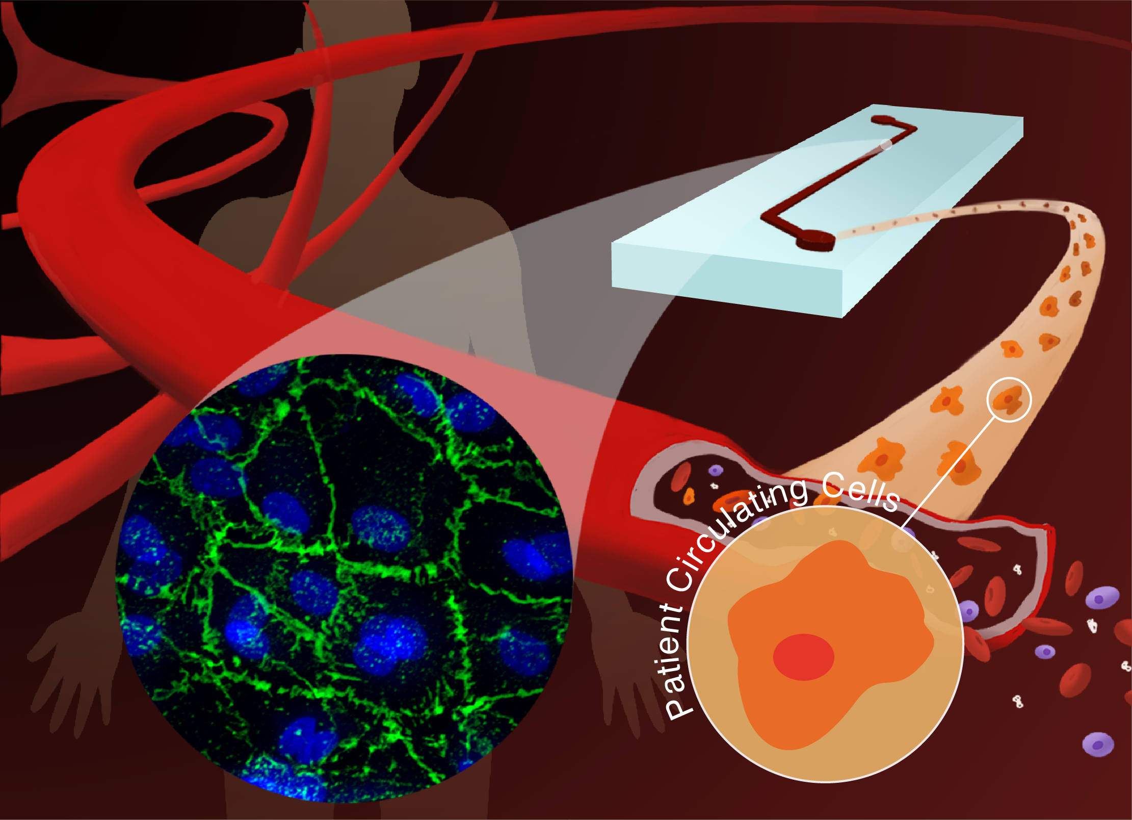
Vascular Organ-Chips made from blood: Enabling personalized medicine of sickle cell disease

In sickle cell disease (SCD), we know that different patients exhibit different extents and frequencies of vaso-occlusive crises. Developing therapeutic strategies against such a diverse phenotype has hence proven to be difficult; the “one-size-fits-all” approach cannot meet the current clinical needs. In my lab, we have demonstrated that easily derived Blood Outgrowth Endothelial Cells (BOECs) from clinically distinct SCD patient blood biopsies can allow visualization of differing patient-phenotypes within the disease through the integration of organ-chip approach with global and single-cell RNAseq. We reveal for the first time that BOEC biopsy is an alternative endothelial cell model for assessing patient-specific regulation of disease severity.

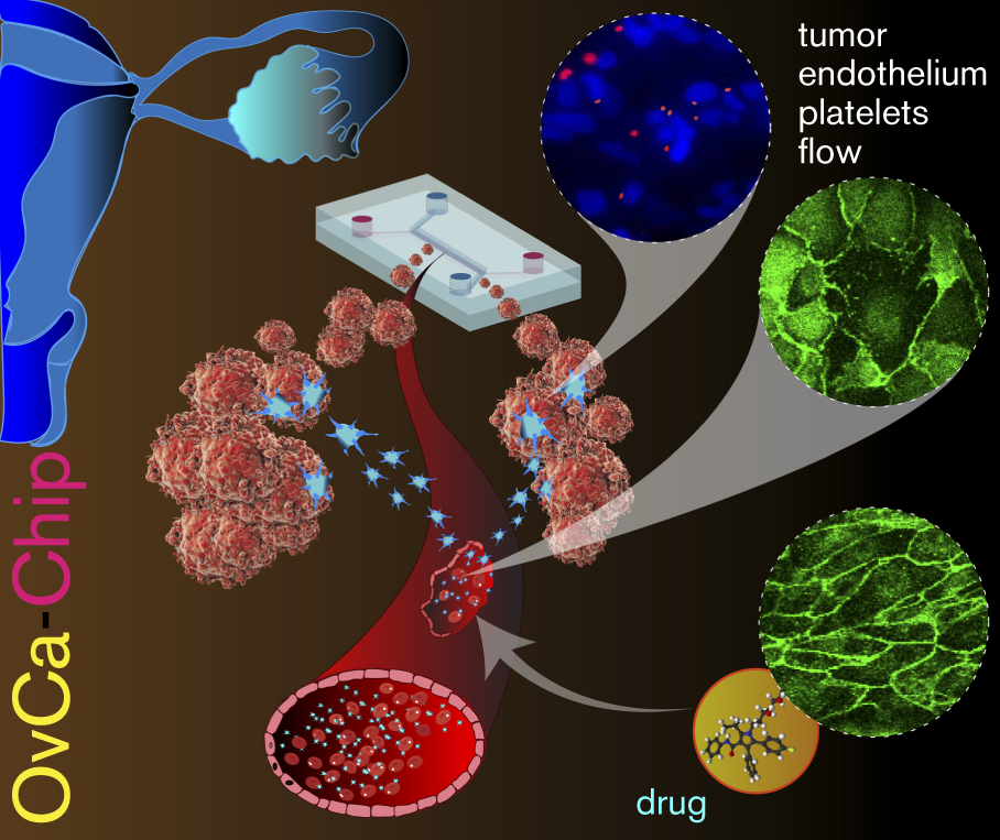
Tumor Microenvironment-Chip: Discovery of platelet immunopathology & combinatorial therapy in cancer

Platelets extravasate from the circulation into tumor microenvironment, enable metastasis and confer resistance to chemotherapy in several cancers. This platelet pathophysiology is suggested to be regulated by the vascular endothelium and its immune function. We have created a new tumor microenvironment-chip (or Tumor-Chip) that recapitulates platelet extravasation through the endothelium and its consequences. By including gene-edited tumors and RNAseq on-chip, this organ-chip revealed temporal dynamics of vascular disintegration due to ovarian cancer cells. We have discovered that platelets and cancer cells interact through glycoprotein GPVI and tumor galectin-3 molecules. As proof-of-principle of a clinical trial, we have revealed that atorvastatin therapy and a novel GPVI inhibitor, Revacept, impairs metastatic potential of cancer and even improves chemotherapy. Since these drugs do not impair hemostasis, our work shows that they are safe cancer therapeutic. Our data are validated by observations in patient tumor samples, and therefore, we propose that our Tumor-Chip is a new platform to model the cancer-vascular-hematology nexus and analyses of potential therapeutics.

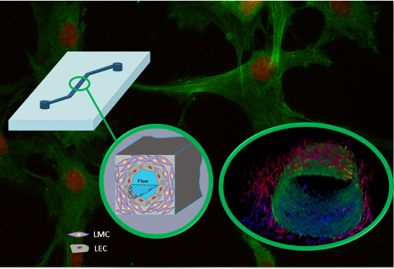
Multicellular Vessel-Chip: Recapitulate hydrodynamics and mechanobiology of lymphedema and atherosclerosis

While the blood vessel is a vast and established theme of research and teaching in medical schools, the lymphatic vessel has traditionally not gained that much attention. As a result, lymphatic diseases, such as lymphedema, still have no cure and there is no dedicated organ-on-chip that models such a disease. We show a new fabrication technique to create a Lymphangion-Chip to model lymphatic physiology under pulsatile flow. This organ-on-chip consists of a co-culture of an endothelial lumen monolayer surrounded by multiple and uniformly thick layers of mural cells. In this device, lymphatic muscle cells align circumferentially while endothelial cells aligned axially under flow, as only observed in vivo in the past. Another effort is to model aging-induced atherosclerosis using iPSC-derived blood vascular endothelial

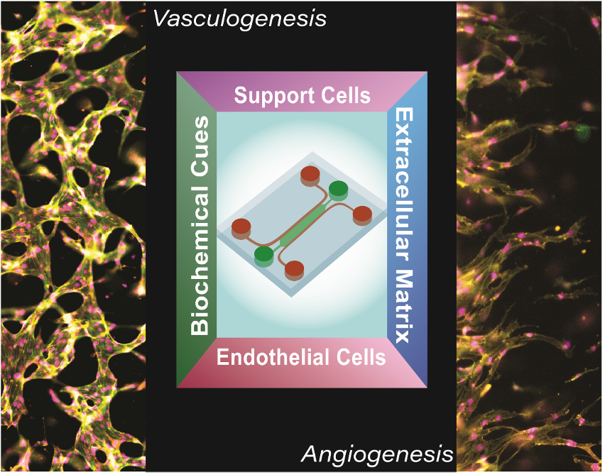
Vascularized Organ-Chips

Almost every organ requires a vasculature for its nutrition and survival, but microvascular networks are rarely incorporated into the organ-chip devices. We are taking a machine learning approach to objectively predict the vasculogenic and angiogenic potential and performance of an organ in a variety of vascularized microphysiological systems. The technique has the potential to break new ground for improving future organ-chips and drug delivery methods.

Microfluidic Devices of Coagulation and Platelet Function

Pediatric patients on mechanical life support systems such as extracorporeal membrane oxygenation (ECMO) and ventricular assist devices (VAD) have a high bleeding and thrombotic risk. Although various coagulation monitoring tests have been developed to guide therapies that minimize these risks, bleeding and thrombosis episodes still occur in patients. A major deficiency with these tests is that they measure clotting characteristics under conditions that are not physiologically relevant, thus limiting the performance of these tests. To address this deficiency, we have designed a microfluidic device that mimics a stenosed, tortuous arteriolar network that permits the flow of whole human blood. By applying a clotting time algorithm to fluid pressure readouts, we have found that this device is sensitive to anticoagulants, platelet concentration, and fibrinolytics.

Department of Biomedical Engineering
Emerging Technologies Building
101 Bizzell St, 3120 TAMU
College Station, TX, 77843

Phone: +1 (979) 458-8494 (office), +1 (979) 847-8113 (lab)

Twitter: JainLabTAMU

© 2016–2023 The Bioinspired Translational Microsystems Laboratory Log in

Texas A&M Engineering Experiment Station Logo